
| About ROSAT |
ROSAT Home Page | ROSAT Images |
|---|


The HRI with its CsI-coated MCPs, was found during ground testing to possess a crude spectral sensitivity which has been verified with in-flight data. Because such a capability lies beyond the baseline HRI requirements, however, resources to permit the quantitative exploitation of this potential were never part of the RSDC budget. Consequently, although we have acquired the necessary data to make this possible, the actual task of fully understanding and calibrating the HRI spectral response has not been completed. Any efforts to derive quantitative spectral results from HRI data must therefore entail extensive, careful analysis on the part of individual investigators.
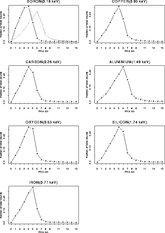
Figure 5.16: The pulse height distribution from ground-based measurements at
7 energies within a ![]() strip centered on the detector.
The dashed curve in the Boron panel is the PHA distribution of
the Al data, and is included to show the full spectral range.
strip centered on the detector.
The dashed curve in the Boron panel is the PHA distribution of
the Al data, and is included to show the full spectral range.
Figure 5.16 shows the PHA distribution from ground-based measurements at 7 energies, and Tab. 5.7 lists the mean PHA channel at each energy. Note that there is a systematic increase in the mean pulse height channel with increasing photon energy except for the Si line (the Si measurement may have been affected by the presence of Si in the glass in the microchannel plates). Table 5.7 shows that the mean PHA varies by approximately 2 channels over the energy range of the ROSAT mirror.